Cutting-edge scanning ion conductance microscopy for the life sciences
Innovative non-invasive nanopipette nanoprobes enable simultaneous acquisition of 3D topography and biochemical imaging of living cells

Yuri Korchev, Professor, Nano Life Sciences Institute (WPI-NanoLSI), Kanazawa University
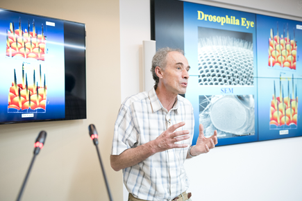
Biophysicist Yuri Korchev is an overseas PI at the Nanometrology group of the WPI NanoLSI and a professor at the Faculty of Medicine, Imperial College London. “I am originally from Ukraine and was given the opportunity to study in the UK and eventually got this position,” says Korchev. “For the WPI project my main input is on scanning ion conductance microscopy imaging of living cells using unique non-contact glass micropipette probes that yield both topographic and chemical images without physically touching cells during scanning in aqueous solutions. We have now extended this research to the fabrication of nanopore extended FETs (nexFET) for selectively sensing proteins.”
Other potential applications of this technology include glucose sensor chips, which could be attached to an individual’s skin for continuous measurements of glucose levels. Another application assisting doctors during cancer surgery to identify cancerous and non-cancerous tissue by testing their chemical properties with the nano sensors.
COVID-19 and research in the UK
Korchev says that the restrictions in the UK are currently much stricter than those in Japan. “In Japan people wear masks and this dramatically reduced the spread of the virus since the early days,” says Korchev. “In contrast official guidelines on wearing mask here were introduced rather late with the result that infection rates are higher than Japan and many other countries in the world.”
Regarding research, Korchev adds that Imperial College was quick to introduce measures to handle the pandemic and that he has been able to continue his research based on the new rules. “It’s a tough schedule where we can visit the labs for 4 days during the week and then we must stay isolated for the next 10 days,” says Korchev. “It is easier for my group because each lab room is separate, so there is little interaction between people.”
Research at NanoLSI
“At the NanoLSI I collaborate with Professor Yasufumi Takahashi who is an expert on scanning electrochemical microscopy,” says Korchev. “I also work with my ex post-doc who recently joined NanoLSI in Kanazawa as an associate professor. We are continuously planning experiments and are proceeding well.” Intriguingly, experiments between the two groups located in London and Kanazawa are completely automated, so scientists share screens of computers that actually control scanning probe measurements at the NanoLSI. This remote-access enables real-time monitoring of experiments by all the participants—an enormously effective means of pursuing international collaborative research under the constraints due to the COVID-19 pandemic.
“The WPI is a very clever idea for pursuing genuine, long term, international, collaborative research,” says Korchev. “The funding enables us to travel to each other’s institutes for significant periods of time. I usually go to Kanazawa about three times a year for periods of around five weeks. I am really forward to visiting Kanazawa again, especially to see the new dedicated NanoLSI building that was opened in December 2020.”
Research highlights
In their seminal 2019 paper, Korchev and his colleagues reported on the potential application of scanning nanopipette scanning ion conductance microscopy for monitoring cancer treatment [1]. They fabricated innovative functionalized nanopipettes incorporating zwitterionic membranes to rapidly monitor changes in extracellular pH levels surrounding living cells; the pH levels are indicative traits of invasive cancer cells and their response to treatment. This system enables measurements of changes in pH of less than 0.01 units, with a response time of 2 ms, and 50 nm spatial resolution.
The research that laid the foundations for this approach to monitoring cancer treatment was reported in two earlier papers. The first in 2016, described the development of “double barrel nanopipette–nanometric field-effect-transistor (FET) sensors with dual carbon nanoelectrodes” as sensors to identify the biochemical properties of single living cells [2].
Then in 2017 Korchev and his colleagues reported the development of a “new class of nanoscale sensors dubbed nanopore extended field-effect transistors (nexFET) that combine the advantages of nanopore single-molecule sensing, field-effect transistors, and recognition chemistry.” The researchers switched gates on and off to control the movement of single biomolecules into the pipette and thereby selectively “sense an anti-insulin in the presence of IgG isotype.” Importantly, incorporating the zwitterionic membrane enabled much faster responses than carbon electrodes as later reported in Nature Communications [1].
References
[1] Y. Zhang et al. High-resolution label-free 3D mapping of extracellular pH of single living cells. Nat Commun 10, 5610 (2019).
https://doi.org/10.1038/s41467-019-13535-1
[2] Y. Zhang et al. Spearhead Nanometric Field-Effect Transistor Sensors for Single-Cell Analysis, ACS Nano. 10(3): 3214–3221, (2016). https://pubs.acs.org/doi/10.1021/acsnano.5b05211
[3] R. Ren, et al. Nanopore extended field-effect transistor for selective single-molecule biosensing. Nat Commun 8, 586 (2017).
https://doi.org/10.1038/s41467-017-00549-w
Further information
NanoLSI Podcasts
This episode offers greater insights into Professor Yuri Korchev’s research at the NanoLSI.


