

Scientists discover how a ring-shaped protein captures and brings together DNA strands
Researchers at Kanazawa University and the National Institute of Genetics in Japan have used high-speed atomic force microscopy to observe how a group of proteins known as Smc5/6 (*1) interacts with DNA (*2). The images reveal how this structure opens and closes, captures DNA and brings separate sections together. The researchers also observed Smc5/6 holding twisted sections of DNA in place, producing more tightly packed arrangements. The findings provide new insights into the molecular processes that contribute to the maintenance and repair of genetic material.
Watching a protein structure change shape
Every living cell must protect and maintain its DNA. This genetic material contains the instructions needed for life, but it is constantly subjected to stresses that can damage its structure. DNA must also be copied accurately when cells divide. To accomplish these tasks, cells rely on proteins that interact with DNA, control its arrangement and support the processes needed to maintain its integrity.
One of these is Smc5/6, a structure made up of several proteins that work together. It belongs to a family of proteins involved in maintaining chromosomes, the structures that carry DNA inside cells. Although scientists have known that Smc5/6 is important for DNA maintenance and repair, precisely how it captures and handles DNA has remained difficult to observe.
The research team used high-speed atomic force microscopy (HS-AFM) (*3), a technique that uses an extremely fine probe to follow movements and structural changes at the nanometre scale. Unlike conventional imaging methods that provide mainly static views, HS-AFM can record sequences of images showing how individual molecular structures behave over time.
The team studied purified Smc5/6 from budding yeast, observing its movements and interactions with DNA under controlled laboratory conditions.
They found that Smc5/6 could adopt two distinct shapes. In one, its two long arms lay close together, giving it an appearance resembling the letter I. In the other, the arms separated to form an open structure resembling the letter O.
The transition between these shapes was influenced by adenosine triphosphate (ATP) (*4), a molecule that supplies energy for many cellular processes. Binding ATP promoted the open configuration, while subsequent ATP-related changes were associated with different ways of holding DNA.
Capturing and bringing DNA together
The images revealed that Smc5/6 could interact with DNA at different parts of its structure. In some configurations, DNA was held near regions at one end of the protein assembly. In others, DNA was positioned within the structure formed by its arms, providing evidence of how Smc5/6 can capture DNA.
The researchers also observed Smc5/6 contacting two separate sections of DNA simultaneously. This allowed the protein assembly to hold the sections together, a process known as DNA tethering.
Another important observation concerned DNA twisting. When Smc5/6 interacted with twisted DNA structures, the DNA occupied a smaller area than DNA without the protein assembly. The results suggest that Smc5/6 can contribute to DNA compaction (*5) by bringing sections of DNA together and stabilizing twisted arrangements.
Understanding how cells maintain their genetic material
The study provides a clearer picture of how Smc5/6 interacts with DNA through coordinated changes in its shape. Rather than simply attaching to DNA at one location, the protein assembly can adopt different configurations that allow it to hold and reposition genetic material.
These properties are relevant to the wider biological roles of Smc5/6, including its involvement in chromosome maintenance and DNA repair.
The experiments were carried out using purified proteins and DNA outside living cells. They therefore reveal molecular interactions under controlled conditions rather than directly showing how the complete process operates inside a cell.
Potential significance
The research advances our understanding of how proteins physically handle DNA. By revealing the movements of Smc5/6 and showing how these movements relate to DNA capture, tethering and compaction, the study provides new information about the molecular mechanisms involved in maintaining chromosomes.
A better understanding of these mechanisms may eventually contribute to research on problems arising from failures in DNA maintenance and repair. However, the present study focuses on fundamental molecular biology and does not demonstrate a medical treatment or clinical application.
“By observing individual Smc5/6 structures as they interacted with DNA, we could see how changes in their shape are associated with different ways of capturing and holding DNA,” says Kenichi Umeda of Kanazawa University. “These observations bring us closer to understanding how this protein assembly carries out its roles in maintaining chromosomes.”
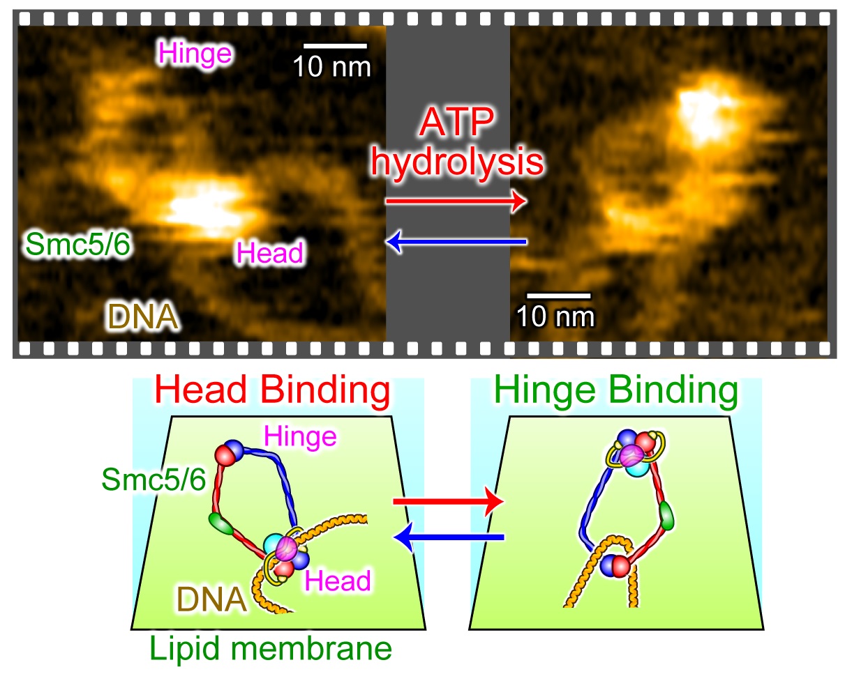
High-speed AFM images of Smc5/6 topologically bound to DNA in the head-binding mode (left) and hinge-binding mode (right).
Study limitations
This study examined purified Smc5/6 and DNA in controlled laboratory experiments. The images do not directly establish every step of Smc5/6 activity inside living cells.
Glossary
Article
- Title
- Submolecular video-imaging of the Smc5/6 complex topologically bound to DNA
- Author
- Kenichi Umeda, Yumiko Kurokawa, Noriyuki Kodera, Yasuto Murayama
- Journal
- Nature Communications
- Publication date
- Sep 1, 2026
- DOI
- 10.1038/s41467-026-77122-x
- URL
- https://doi.org/10.1038/s41467-026-77122-x