Flexible motion of bacteriophage tail fibers modulates infection
In this study, as part of an international collaboration supported in part by the Bio-SPM Summer School and the Collaborative Research Program (2024–2025), a research team led by Associate Professor Hiroki Konno and Soma Yamamoto (master’s student in the Division of Life Science and Engineering and member of the WISE Program for Nano-Precision Medicine, Science, and Engineering) at the Nano Life Science Institute (WPI-NanoLSI), Kanazawa University, together with Professor Noriyuki Kodera (WPI-NanoLSI), in collaboration with Assistant Professor Balint Kiss and Professor Miklos Kellermayer (Department of Biophysics and Radiation Biology, Semmelweis University, Hungary), has successfully elucidated a key dynamic mechanism utilized by bacteriophages in the process of recognizing the surface of their host (bacterial) cells.
This study focuses on the tail fibers of T7 bacteriophage (*1), which were expressed and purified at the Department of Biophysics and Radiation Biology, Semmelweis University (Budapest, Hungary). The fibers were subsequently imaged using high-speed atomic force microscopy (HS-AFM) at the Nano Life Science Institute (WPI-NanoLSI), Kanazawa University (Kanazawa, Japan).
This study highlights the crucial role of viral protein dynamics in the T7 bacteriophage infection process. HS-AFM observations reveal that T7 tail fibers exhibit both bending and torsional flexibility, suggesting that these mechanical properties are actively utilized by the phage during host cell surface exploration.
The results were published online as Open Access in Small (Wiley) on June 19, 2026.
【Background】
Bacteriophages—viruses that specifically infect bacteria—have attracted increasing attention in recent years due to their potential therapeutic applications in overcoming multidrug-resistant bacterial infections.
In this study, the team investigated the structural dynamics of the tail fibers of T7 bacteriophage. The T7 virion consists of an icosahedral capsid (*2) and a tail-fiber complex, which plays a critical role in bacterial target recognition and viral DNA injection. The virus possesses six L-shaped fibers, approximately 40 nm in length, attached to the tail tube. These fibers are believed to be essential for initial host recognition and may also facilitate surface exploration.
During the first step of infection, phages recognize and attach to the bacterial outer membrane via their tail fibers. This is followed by full phage immobilization through engagement of the tail tube. However, the precise sequence of events and the mechanisms triggering these binding processes remain unclear.
【Summary of the Research Findings】
Using high-speed atomic force microscopy (HS-AFM), molecular dynamics (MD) simulations, and small-angle X-ray scattering (SAXS), the team analyzed the molecular structure and dynamics of isolated tail fibers and tail-fiber complexes (Fig. 1).
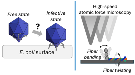
Fig. 1. During host recognition, T7 bacteriophage fibers transition from a capsid-bound state to an extended conformation, a process that requires structural flexibility (left). HS-AFM observation demonstrates the fibers contain a molecular hinge and a torsionally compliant coiled-coil region (right).
The results reveal that the kink region separating the proximal and distal segments of the fiber functions as a molecular hinge, enabling large-scale bending motions. This flexibility may enable the phage to explore the host surface through a “walking-like” mechanism.
Additionally, partial unwinding and rewinding within the triple-helical coiled-coil structure (*3) of the proximal segment allow for rotational and torsional flexibility. These dynamic features enable rapid, large-scale flexing and extension of the fibers, facilitating an efficient topological search for anchoring sites on the host cell surface.
Because fiber-assisted topological search is likely a general mechanism of host recognition, its modulation may provide a means to fine-tune phage–bacterium interactions.
【Future Perspectives】
This study characterizes the large-scale motions of T7 tail fibers and suggests their functional role in host cell surface exploration. As a next step, the team aims to directly observe the movement of tail-fiber complexes on the surface of living bacteria using HS-AFM.
In addition, the team plans to investigate the nanomechanics of fiber–host binding using optical tweezers, thereby providing further insight into the physical forces underlying the infection process.
Glossary
Article
- Title
- Large-Scale Structural Dynamics in the Tail Fiber Modulate the Infective Transition of the T7 Bacteriophage.
- Author
- Luca Elizabet Kosik, Miklós Cervenak, Dominik Sziklai, Andrea Balogh-Molnár, Negar Rahimi, Bence Fehér, Soma Yamamoto, Hiroki Konno, Noriyuki Kodera, Holger Flechsig, Romain Amyot, Heinz Amenitsch, Hedvig Tordai, Levente Herényi, Ana Cuervo, Miklós Kellermayer, Bálint Kiss.
- Journal
- Small
- Publication date
- Jun 19, 2026
- DOI
- 10.1002/smll.74269
- URL
- https://onlinelibrary.wiley.com/doi/10.1002/smll.74269