Elucidation of progression mechanism of glioma mediated by extracellular vesicles
Abstract
A research group from the Nano Life Science Institute, Kanazawa University, led by Profs. Rikinari Hanayama and Hironori Kawahara in collaboration with a group from Kanazawa University Graduate School of Medicine led by Profs. Mitsutoshi Nakada and Taishi Tsutsui has succeeded in elucidating the mechanism of glioma progression including invasion and metastasis by extracellular vesicles (EVs). Glioma are among the most aggressive primary tumors of the central nervous system; prognosis is poor with a median survival time of about 2 years even with multimodal therapy. The scientists have revealed that glioma-derived EVs convey a tumor-related factor WT1 to microglial cells in the surrounding areas of a tumor leading to angiogenesis (generation of new blood vessels), which contributes to the creation of a microenvironment favorable for invasion and metastasis of a tumor.
Using an intracranial implantation glioma mouse model, they first showed a longer survival time of mice through tumor size reduction and suppression of invasion and metastasis by repressing the production of EVs by the glioma. Next, they showed that EVs secreted from the glioma were taken up by the surrounding microglia thus regulating gene expression in microglia, thereby promoting angiogenesis. Furthermore, they showed that WT1, an expression regulator of that gene, was contained in tumor-derived EVs of human patients and regulated the microglial ability of angiogenesis. This study indicates that EVs are deeply involved in the progression of glioma and it is suggested that repression of their production may suppress tumor invasion and metastasis. It is expected that this research will lead to the early detection and prognosis of glioma and the development of new treatments in the near future.
Background
In recent years, research on extracellular vesicles (EVs)*1) has been developed in various fields of medical research such as immunology, neurology and cancer. EVs are tiny endogenous particles secreted by almost all cells in the body. They contain proteins, nucleic acids, lipids, etc. specific to the secreting cells. Since these constituents differ depending on the secreting cells and diseases, EVs from body fluids such as blood and urine are expected to be useful as biomarkers for the early detection and prognosis of diseases. It has also been shown that EVs induce various cellular responses by delivering these molecules to surrounding cells and are involved in the development of various life phenomena and diseases. The scientists from Kanazawa University investigated the involvement of tumor-derived EVs in the progression of glioma. Glioma are known to be among the most aggressive primary tumors of the brain; prognosis is poor with a median survival time of about 2 years even with multimodal treatment combining surgery, radiation therapy and anticancer drug treatment. Therefore, urgent elucidation of the progression mechanism and development of new therapeutic methods are clearly needed.
Outline of research results
The scientists from Kanazawa University have established a glioma cell line deficient in a molecule involved in producing EVs and have generated a glioma mouse model by implanting the cell line into the brain. By using these mice, it was revealed that repressing EV production reduces glioma size to 1/10 or less, suppresses invasion and metastasis in the brain, and prolongs mouse survival time by 25% (Figure 1). Next, they found that EVs secreted by glioma are ingested by microglia*2) in the surrounding areas of the tumor where they reduce microglial gene expression of thrombospondin*3), an inhibitory factor of angiogenesis, resulting in enhanced angiogenesis mediated by microglia. Furthermore, they showed that WT1*4), a downregulator of the thrombospondin gene, was contained in the tumor-derived EVs of human patients of glioma and upregulated the ability for angiogenesis of microglia (Figure 2).
Future prospect
The present study has revealed, through indicating that EVs secreted by glioma enhance angiogenesis via microglia, the molecular mechanism involved in the establishment of tumor microenvironment*5) promoting tumor invasion and metastasis. It is anticipated that early detection and prognosis of glioma should become possible by quantifying WT1 in EVs involved in the regulation of angiogenesis. Furthermore, development of methods to reduce EV production and WT1 protein levels is expected to lead to new therapeutics for glioma.
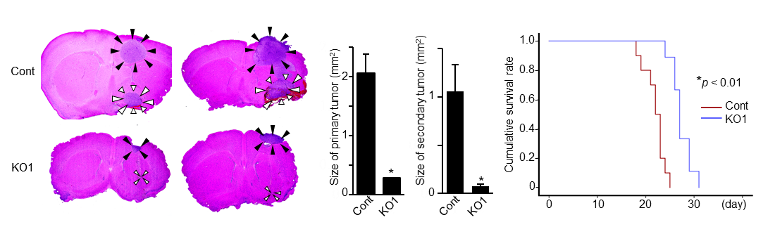
Figure 1. Relationship of extracellular vesicle (EV) production to the invasion and metastasis of glioma.
A glioma cell line (KO1) whose production of EVs is attenuated was established and the progression ability of KO1-derived tumor was compared with that of control cell line-derived tumor (Cont). In the mouse brain, reduction of tumor size (left) and suppression of invasion/metastasis (middle) were observed in KO1-derived tumors and a significant difference in cumulative survival rates of mice was discerned (right).
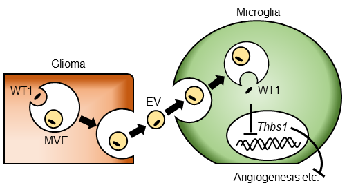
Figure 2. A schematic drawing of angiogenesis regulation by EVs.
Inside multivesicular endosomes (MVE) of glioma cells, WT1 is encapsulated when EVs are formed. The EVs incorporated into microglia release WT1 inside microglia, thus repressing expression of the gene for thrombospondin (Thbs1) that suppresses angiogenesis. As a result, angiogenesis by microglia is enhanced and invasion and metastasis of a tumor are enhanced.
Glossary
Article
- Title
- Glioma-derived extracellular vesicles promote tumor progression by conveying WT1
- Author
- Taishi TSUTSUI, Hironori KAWAHARA, Ryouken KIMURA, Yu DONG, Shabierjiang JIAPAER, Hemragul SABIT, Jiakang ZHANG, Takeshi YOSHIDA, Mitsutoshi NAKADA & Rikinari HANAYAMA
- Journal
- Carcinogenesis
- Publication date
- May 28, 2020
- DOI
- 10.1093/carcin/bgaa052
- URL
- https://europepmc.org/article/med/32463428