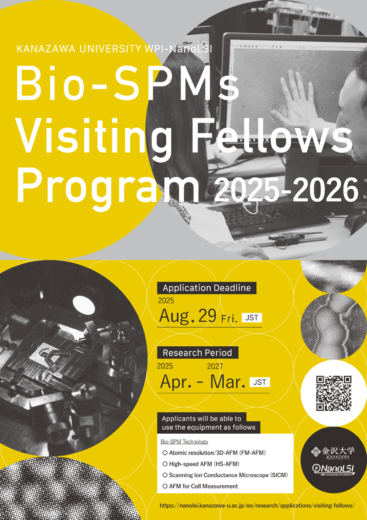
The Nano Life Science Institute (WPI-NanoLSI), Kanazawa University, is calling for applications for NanoLSI Visiting Fellows Program 2025-2026.
 Nano Life Science Institute, Kanazawa University, Japan was chosen on October 6, 2017 to become another center in MEXT’s World Premier International Research Center Initiatives (WPI Centers). http://www.jsps.go.jp/english/e-toplevel/
Head of the Institute, Prof. Takeshi Fukuma, developed the world’s first liquid-environment frequency modulation atomic force microscope (FM-AFM) with true atomic resolution, making it possible to observe the surface structures of biomolecules, and three-dimensional distributions of hydration and flexible surface structures with subnanometer resolution at solid-liquid interfaces. One of the PIs of our WPI project, Prof. Toshio Ando developed high-speed atomic force microscopy (HS-AFM). His team has been carrying out biophysical studies of proteins by observing molecules in action with HS-AFM.
Based on these advanced technologies, NanoLSI plans to develop “nanoendoscopic techniques”. We will combine the world’s most advanced bio-scanning probe microscopy (SPM) and supramolecular chemistry. This combination will allow for not only the imaging of surfaces and interior of live cells but also the analysis of metabolites and nucleic acids and the manipulation of cell activities. By introducing multi-scale simulations to these studies, we aim to construct models for the mechanisms underlying molecular and cellular functions. We also aim to understand cancer-specific abnormalities of cells by comparing normal and cancer cells. Through these studies, NanoLSI aims to achieve nano- level understandings of various life phenomena and thereby establish a new research filed termed “nanoprobe life science”.
Within this framework of NanoLSI’s missions, we activate national and internationalcollaborations. To this end and for the purpose of disseminating our SPM techniques, we have founded NanoLSI Visiting Fellows Program. Under this program, we will invite researchers (PIs) of molecular, cellular, or structural biologists including sabbatical researchers. For a month or a few months, invited researchers will carry out research, participate in collaborations and other academic activities at NanoLSI, and thereby, catalyze international scientific cooperation between their institutions and NanoLSI.
Invited researchers can learn how to operate the FM-AFM and HS-AFM systems from experts and then perform imaging experiments by themselves on the proteins and cells that they are willing to study.
The NanoLSI Visiting Fellows Program covers:
- Travel expenses
- Accommodation expenses and daily allowance based on the KU regulations
In addition that,
- Applicants might be accompanied by researchers, postdoc, etc. in his/her own lab. NanoLSI will also cover maximum two researchers’ travel expenses.
- Faculty members and technicians fully support applicants’ research activities at NanoLSI.
- While staying at NanoLSI, applicants and accompanying persons will be able to use the whole laboratory, including instruments, supplies (up to 300K JPY), etc.
- During the research stay, applicants and accompanying persons can stay in a university guest house (about 3,000 JPY per night) if there is a vacancy.
Applicants will be able to use the following instruments.
Atomic resolution AFM (FM-AFM & 3D-AFM)
FM-AFM (Frequency-Modulation Atomic Force Microscope) can visualize subnanometer-scale surface structures of biomolecules in solution. Combined with 3D scanning technique, it can also visualize 3D distribution of hydration and flexible surface structures at solid-liquid interfaces. The imaging rate of FM-AFM and 3D-AFM is typically 1 min/frame. The optimal spatial resolution of the instrument is 0.3 nm in the lateral direction and 0.01 nm in the vertical direction. In the case of biomolecular imaging, the practical resolution is mostly determined by the fluctuation of the surface structures rather than the instruments. For more details, see the following articles:
- H. Asakawa, S. Yoshioka, K. Nishimura, T. Fukuma, “Spatial Distribution of Lipid Headgroups and Water Molecules at Membrane/Water Interfaces Visualized by Three-Dimensional Scanning Force Microscopy”, ACS Nano 6, 9013-9020 (2012).
- A. Yurtsever, H. Asakawa, Y. Katagiri, K. Takao, K. Ikegami, M. Tsukada, M. Setou, T. Fukuma, “Visualizing the Submolecular Organization of alphabeta-Tubulin Subunits on the Microtubule Inner Surface Using Atomic Force Microscopy.” Nano Lett, 25, 98-105 (2025).
- T. Fukuma, R Garcia, “Atomic- and Molecular-Resolution Mapping of Solid-Liquid Interfaces by 3D Atomic Force Microscopy.” ACS Nano, 12 (12), 11785-11797 (2018).
High-Speed AFM (HS-AFM)
HS-AFM (High-Speed Atomic Force Microscope) can visualize moving objects in solution. Its temporal resolution is typically 100 ms/frame, while the spatial resolution is 2-3 nm in the lateral direction and 0.15 nm in the vertical direction. When it is applied to protein molecules in action, the acquired HS-AFM images can provide a significant insight into how the molecules function. For more details, see the following review articles:
- T. Ando, T. Uchihashi, S. Scheuring, “Filming biomolecular processes by high-speed atomic force microscopy”, Chem. Rev. 114, 3120-3188 (2014).
- T. Ando, S. Fukuda, K. X. Ngo, “High-Speed Atomic Force Microscopy for Filming Protein Molecules in Dynamic Action”, Annu. Rev. Biophys. 53, 19-39 (2024).
- T. Uchihashi, N. Kodera, T. Ando, “Guide to video recording of structure dynamics and dynamic processes of proteins by high-speed atomic force microscopy”, Nature Protocols 7, 1193-1206 (2012).
Scanning Ion Conductance Microscopy (SICM)
SICM has a unique measurement principle and provides an unprecedented opportunity that enables submicroscale functional imaging of single live cells by a combination of nanoscale local stimulation and noncontact topography imaging. The imaging rate of SICM is 30-300 s/frame. Spatial resolution of the instrument is 10 nm in the lateral direction and 5 nm in the vertical direction. For more details, see the following articles:
- P. Novak, C. Li, A. I. Shevchuk, R. Stepanyan, M. Caldwell, S. Hughes, T. G. Smart, J. Gorelik, V. P. Ostanin, M. J. Lab, G. W. J. Moss, G. I. Frolenkov, D. Klenerman, and Y. E. Korchev, “Nanoscale live-cell imaging using hopping probe ion conductance microscopy”, Nat. Methods 6, 279-281 (2009).
- V. O. Nikolaev, A. Moshkov, A. R. Lyon, M. Miragoli, P. Novak, H. Paur, M. J. Lohse, Y. E. Korchev, S. E. Harding, and J. Gorelik, “beta(2)-Adrenergic Receptor Redistribution in Heart Failure Changes cAMP Compartmentation”, Science 327, 1653-1657 (2010).
- Zhou, M. Saito, T. Miyamoto, P. Novak, A. Shevchuk, Y. Korchev, T. Fukuma, Y. Takahashi, “Nanoscale Imaging of Primary Cilia with Scanning Ion Conductance Microscopy,” Anal. Chem. 90, 2891-2895 (2018).
AFM for Cell Measurement
Based on high-speed AFM or 3D-AFM, NanoLSI is developing AFM technologies for measuring the structure, dynamics or mechanical properties of the surface or inside of cells at a nano scale. High-speed AFM successfully visualized the surface structure of bacteria at a molecular scale and nano-motion of the terminal portion of nerve cells. Based on 3D-AFM, we developed a nanoendoscopy-AFM technique. Using this technique, we succeeded in three-dimensional observation of cell nucleus or actin fibers inside live cells, the measurement of two-dimensional nanodynamics of inner scaffold of plasma membrane, and the measurement of the surface stiffness of cell nucleus. For more details, see the following articles:
- H. Yamashita, A. Taoka; T. Uchihashi, T. Asano, T. Ando, Y. Fukumori, “Single-molecule imaging on living bacterial cell surface by high-speed AFM”, J. Mol. Biol. 422 (2), 300-9 (2012).
- M. Shibata, T. Uchihashi, T. Ando, R. Yasuda, “Long-tip high-speed atomic force microscopy for nanometer-scale imaging in live cells”, Sci. Rep., 5, 8724 (2015).
- M. Penedo, K. Miyazawa, N. Okano, Furusho, H. Ichikawa, T, S. Alam Mohammad, K. Miyata, C. Nakamura, T. Fukuma, “Visualizing intracellular nanostructures of living cells by nanoendoscopy-AFM”, Sci. Adv. 7 (52), eabj4990 (2021).
- K. Kobayashi, N. Kodera, T. Kasai, YO. Tahara, T. Toyonaga, M. Mizutani, I. Fujiwara, T. Ando, M. Miyata. “Movements of Mycoplasma mobile gliding machinery detected by high-speed atomic force microscopy”, mBio 12: e00040-21 (2021).
Application Eligibility
Applicants must be independent researchers who lead an independent group or run their own lab.
Application Procedure
Applicants should submit:
Application Deadline
Friday, August 29, 2025 Japan time
Selection and Result
Based on the submitted documents above, the Selection Committee will select and nominate candidates to the Head of the Institute. The notification will be sent to the applicants in October 2025 .
For further information, please contact:
Yuki Kunioka, Ph.D.
Research Administrator
Nano Life Science Institute, Kanazawa University
Kakuma, Kanazawa, Ishikawa 920-1192 JAPAN
nanolsi_openf01@ml.kanazawa-u.ac.jp

