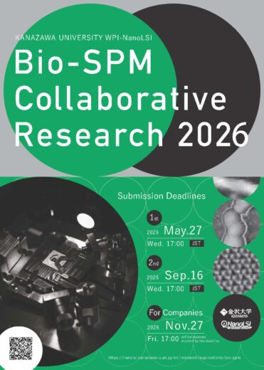
Call for Bio-SPM Collaborative Research Proposal


I. Outline of Call for Research Proposals

1. Aims

The research teams at Kanazawa University have engaged in the pioneering development of original Bio-SPM technologies, including Atomic resolution/3D-AFM (FM-AFM), High-speed AFM, Scanning Ion Conductance Microscope (SICM), and AFM for Cell Measurement, and applied them to the life sciences. Here, we call for applications for collaborative research projects that are carried out by the applicants using our Bio-SPMs, with the cooperation of the NanoLSI faculty members.

2. Type of Collaborative Research

1) Collaborative Research by visits to NanoLSI
Applicants adopted visit WPI-NanoLSI and carry out a research project, with the cooperation of the NanoLSI faculty members.

2) Collaborative Research by sending samples
Applicants adopted do not visit WPI-NanoLSI. Samples are sent to WPI-NanoLSI and researchers in charge at WPI-NanoLSI conduct experiments. Please note that this option is limited to cases where visiting WPI-NanoLSI is difficult due to COVID-19-related issues. In addition, the applicant is supposed to obtain permission from the researcher in charge before application.

3. Application Eligibility

Researchers or engineers from national and overseas public or private universities.

Note:

Students such as graduate students and undergraduate students are not eligible as an applicant but can be included in the research project team.

  • “NanoLSI Associates” are accepted to apply even if they belong to companies.
  • For companies, please see this (in preparation).

4.  Research Period

For applicants adopted in 1st or 2nd call, from the date of approval to March 31, 2024.
For applicants adopted in 3rd call, research can be carried out after March 31, 2024

Note:

You are supposed to inform a research period within one month after the notification of approval. We may not be able to meet your request for the timing of collaboration due to circumstances.

5.  Decisions

After the review by the Expert Committee of NanoLSI, Kanazawa University, the director of NanoLSI will decide which applications will be approved. The decision will be informed to the applicants.

6. How to Apply

Fill out Application Form 1 and submit it by the following deadline.

Form 1  “2023 Academic Year Bio-SPMs Collaborative Research, Application and Collaborative Researcher Approval Form”  PDF  Word

Arbitrary format “Curriculum vitae for the principal investigator (applicant) only”

Submission deadlines

1st in 2023:  May 31, 2023 (Wednesday) 17:00 JST
2nd in 2023:  July 31, 2023 (Monday) 17:00 JST
3rd in 2023:  October 30, 2023 (Monday) 17:00 JST

Note:

For urgent cases, applications will be accepted regardless of the deadline. However, this procedure is only available after coordination with the researchers in the institute.

Submission Destination

Nano Life Science Institute, Kanazawa University,
Kakuma-Machi, Kanazawa, 920-1192, Japan.
E-mail:  nanolsi_openf01*ml.kanazawa-u.ac.jp
Please replace the asterisks (*) with @.

7. Inquiries Regarding Research Contents

Send an inquiry to the e-mail address below. The inquiry should include the type of Bio-SPMs you wish to use (either Super-resolution AFM [FM-AFM/3D-AFM], High-speed AFM, SICM, or AFM for cell measurement). The reply will come from the person in charge of the specific Bio-SPM technology.

E-mail:  nanolsi_openf01*ml.kanazawa-u.ac.jp
Please replace the asterisks (*) with @.

II. Post-Acceptance Procedures

8. Research Report

An approved researcher has an obligation to report the research results when the research period is over. Using Forms 2 and 3, prepare and submit the research report. The deadline is May 10, 2024. The summary of research results (Form 3) will be publicly posted on the NanoLSI Bio-SPMs Collaborative Research website in the 2023 academic year.

(In preparation)

Form 2: “2023 Academic Year Bio-SPMs Collaborative Research, Research Report” PDF Word
Form 3: “2023 Academic Year Bio-SPMs Collaborative Research, Research Report Summary” PDF Word

9.  When the Research Papers are Published

In order to measure the effectiveness of this Bio-SPMs collaborative research activity, we would like to collect the data of research papers published through collaborative research carried out at NanoLSI. Therefore, we sincerely hope that the approved researchers will kindly inform us when the research papers are published. In addition, for three years after the research period is over, we would like to ask all the approved researchers every June via e-mail if the research papers through collaborative research were newly published.

Supplementary Information
  • Regarding the Birth date, Age, and Gender fields in the Application
    These fields are necessary for making statistics for the evaluation report by the Ministry of Education, Culture, Sports, Science and Technology, in which the ages and genders of collaborative researchers must be presented. The information provided will not influence any decisions. In addition, the personal information provided will be handled with the utmost caution.
  • Regarding Filling out the Application
    Add more spaces freely if not enough space is available in the application form. The number of pages can be increased.

III.  Others

  1. Research applications that match our policy described in “1. Aims” will be preferentially approved. It is preferable that applicants have already conducted sample preparations and preliminary experiments, for example, biochemical experiments, tests of observation conditions, and microscopic analysis by optical microscope, electron microscope, SPM, etc. However, applications without any preliminary results will also be approved depending on the contents.
  2. If your samples or methods are quite new to Bio-SPM measurements, we will select your proposal as a “Preliminary Investigation”. In Preliminary Investigation, Experts of Bio-SPM will perform some experiments to search for the measurement conditions. When the measurement conditions for your proposal are fixed, you can start full research.
  3. When a submitted application is approved, the principal investigator (PI) responsible for the collaborative research and the collaborative partner researcher should be a collaborative researcher of NanoLSI, Kanazawa University. All members who participate in the approved research project can stay at Kakuma Guest House (http://guesthouse.w3.kanazawa-u.ac.jp/index.html). If no room is available at Kakuma Guest House, stay at hotels downtown.
  4. If it is necessary to support the travel expenses including transportation and accommodation for staying at Kanazawa University, mention it in the appropriate part of Form 1. Travel expenses will be supported by up to 200K JPY for domestic applicants and 350K JPY for overseas applicants based on the travel expense regulations of Kanazawa University after the review by the Expert Committee of NanoLSI. (However, if the airfare is very expensive, WPI-NanoLSI may support up to one person’s round-trip travel expenses plus one week’s accommodation expenses, exceeding the maximum limit.) In the case of collaborative research by sending samples, WPI-NanoLSI will bear the shipping costs for the samples without travel fees, however, will not be covered travel expenses.
  5. For the outstanding proposals related to cancer research, travel expenses will be supported by up to 200K JPY for domestic applicants and 350K JPY for overseas applicants by the Cancer Research Institute, Kanazawa University based on the travel expense regulations of Kanazawa University. Adopted should mention as follows when the results are published, “This work was partly supported by Extramural Collaborative Research Grant of Cancer Research Institute, Kanazawa University.”
  6. Graduate and undergraduate students can also participate in a collaborative research team. In such cases, approval from their supervisor is absolutely necessary. The approval form is available in Form 1: “2023 Academic Year Bio-SPMs Collaborative Research, Application and Collaborative Researcher Approval Form.” Travel expenses can be covered for only graduate students, but not for undergraduate students.
  7. If the supervisor of the student collaborative researchers changes, new approval from the new supervisor would be necessary. In such cases, contact the person in charge of the Bio-SPMs Collaborative Research (E-mail:  nanolsi_openf01*ml.kanazawa-u.ac.jp, Please replace the asterisks (*) with @.).
Supplementary Information
  • Regarding the Birth date, Age, and Gender fields in the Application
    These fields are necessary for producing statistics for the evaluation report by the Ministry of Education, Culture, Sports, Science and Technology, in which the ages and genders of collaborative researchers must be presented. The information provided will not influence any decisions. In addition, the personal information provided will be handled with the utmost caution.
  • Regarding Filling out the Application
    Add more spaces freely if not enough space is available in the application form. The number of pages can be increased.

Overview of each Bio-SPM Technology

Atomic resolution AFM (FM-AFM & 3D-AFM)

FM-AFM (Frequency-modulation Atomic Force Microscope)can visualize subnanometer-scale surface structures of biomolecules in solution. Combined with 3D scanning technique, it can also visualize 3D distribution of hydration and flexible surface structures at solid-liquid interfaces. The imaging rate of FM-AFM and 3D-AFM is typically 1 min/frame. The optimal spatial resolution of the instrument is 0.3 nm in the lateral direction and 0.01 nm in the vertical direction. In the case of biomolecular imaging, the practical resolution is mostly determined by the fluctuation of the surface structures rather than the instruments. For more details, see the following articles:

  1. H. Asakawa, S. Yoshioka, K. Nishimura, T. Fukuma, “Spatial Distribution of Lipid Headgroups and Water Molecules at Membrane/Water Interfaces Visualized by Three-Dimensional Scanning Force Microscopy”, ACS Nano 6, 9013-9020 (2012).
  2. H. Asakawa, K. Ikegami, M. Setou, N. Watanabe, M. Tsukada, T. Fukuma, “Submolecular-Scale Imaging of α-Helices and C-Terminal Domains of Tubulins by Frequency Modulation Atomic Force Microscopy in Liquid”, Biophys. J. 101, 1270-1276 (2011).
  3. T. Fukuma, “Water distribution at solid/liquid interfaces visualized by frequency modulation atomic force microscopy”, Sci. Technol. Adv. Mater. 11, 033003 (18 pages) (2010).

High-speed AFM (HS-AFM)

HS-AFM (High-speed Atomic Force Microscope) can visualize moving objects in solution. Its temporal resolution is typically 100 ms/frame, while the spatial resolution is 2-3 nm in the lateral direction and 0.15 nm in the vertical direction. When it is applied to protein molecules in action, the acquired HS-AFM images can provide significant insight into how the molecules function. For more details, see the following review articles:

  1. T. Ando, T. Uchihashi, S. Scheuring, “Filming biomolecular processes by high-speed atomic force microscopy”, Chem. Rev. 114, 3120-3188 (2014).
  2. T. Ando, T. Uchihashi, N. Kodera, “High-speed AFM and applications to biomolecular systems”, Annu. Rev. Biophys. 42, 393-414 (2013).
  3. T. Uchihashi, N. Kodera, T. Ando, “Guide to video recording of structure dynamics and dynamic processes of proteins by high-speed atomic force microscopy”, Nature Protocols 7, 1193-1206 (2012).

Scanning Ion Conductance Microscopy (SICM)

SICM has a unique measurement principle and provides unprecedented opportunity that enables submicroscale functional imaging of single live cells by a combination of nanoscale local stimulation and noncontact topography imaging. The imaging rate of SICM is 30-300 s/frame. Spatial resolution of the instrument is 10 nm in the lateral direction and 5 nm in the vertical direction. For more details, see the following articles:

  1. P. Novak, C. Li, A. I. Shevchuk, R. Stepanyan, M. Caldwell, S. Hughes, T. G. Smart, J. Gorelik, V. P. Ostanin, M. J. Lab, G. W. J. Moss, G. I. Frolenkov, D. Klenerman, and Y. E. Korchev, “Nanoscale live-cell imaging using hopping probe ion conductance microscopy”, Nat. Methods 6, 279-281 (2009).
  2. V. O. Nikolaev, A. Moshkov, A. R. Lyon, M. Miragoli, P. Novak, H. Paur, M. J. Lohse, Y. E. Korchev, S. E. Harding, and J. Gorelik, “beta(2)-Adrenergic Receptor Redistribution in Heart Failure Changes cAMP Compartmentation”, Science 327, 1653-1657 (2010).
  3. Zhou, M. Saito, T. Miyamoto, P. Novak, A. Shevchuk, Y. Korchev, T. Fukuma, Y. Takahashi, “Nanoscale Imaging of Primary Cilia with Scanning Ion Conductance Microscopy,” Anal. Chem. 90, 2891-2895 (2018).

AFM for Cell Measurement

Based on high-speed AFM or 3D-AFM, NanoLSI is developing AFM technologies for measuring the structure, dynamics or mechanical properties of the surface or inside of cells at a nano scale. High-speed AFM successfully visualized the surface structure of bacteria at a molecular scale and nano-motion of the terminal portion of nerve cells. Based on 3D-AFM, we developed a nanoendoscopy-AFM technique. Using this technique, we succeeded in three-dimensional observation of cell nucleus or actin fibers inside live cells, the measurement of two-dimensional nanodynamics of inner scaffold of plasma membrane, and the measurement of the surface stiffness of cell nucleus. For more details, see the following articles:

  1. H. Yamashita, A. Taoka; T. Uchihashi, T. Asano, T. Ando, Y. Fukumori, “Single-molecule imaging on living bacterial cell surface by high-speed AFM”, J. Mol. Biol. 422 (2), 300-9 (2012).
  2. M. Shibata, T. Uchihashi, T. Ando, R. Yasuda, “Long-tip high-speed atomic force microscopy for nanometer-scale imaging in live cells”, Sci. Rep., 5, 8724 (2015).
  3. M. Penedo, K. Miyazawa, N. Okano, Furusho, H. Ichikawa, T, S. Alam Mohammad, K. Miyata, C. Nakamura, T. Fukuma, “Visualizing intracellular nanostructures of living cells by nanoendoscopy-AFM”, Sci. Adv. 7 (52), eabj4990 (2021).
  4. K. Kobayashi, N. Kodera, T. Kasai, YO. Tahara, T. Toyonaga, M. Mizutani, I. Fujiwara, T. Ando, M. Miyata. “Movements of Mycoplasma mobile gliding machinery detected by high-speed atomic force microscopy”, mBio 12: e00040-21 (2021).

Application Materials

Application Guidelines

Form 1: “2023 Academic Year Bio-SPMs Collaborative Research, Application and Collaborative Researcher Approval Form” PDF Word
Form 2: “2023 Academic Year Bio-SPMs Collaborative Research, Research Report” (In preparation)    
Form 3: “2023 Academic Year Bio-SPMs Collaborative Research, Research Report Summary” (In preparation)    

An approved researcher has an obligation to report the research results when the research period is over. The deadline is May 10, 2024.


For the Reports FY2022, click here.


Updated on 12 June 2023.