Visualizing atomic scale structures and dynamics at solid-liquid interfaces by atomic force microscopy

Kazuki Miyata, Assistant Professor, Nano Life Science Institute (WPI-NanoLSI), Kanazawa University
Focus on solid-liquid interfaces
A deep understanding of the interactions between differing atomic species of materials at the interfaces of solids and liquids plays a major role in the infrastructure of modern society. For example, the key-technology used for the development of water proof clothes, self-cleaning building materials for housing, and protective coatings for car bodies is based on atomic scale insights into the properties of solid-liquid interfaces. But there are still many material systems and phenomena that are not fully understood: the physical chemistry of hydration, crystal growth, and self assembly; corrosion and lubrication for industrial applications; and cell membranes and ion transportation in the life sciences. With this background, Kazuki Miyata is developing unique atomic force microscopy systems (AFM) that are capable of imaging solid-liquid interfaces of a wide range of materials. “The scanning tunneling microscope, stimulated emission depletion microscopy and cryo-electron microscopy are inventions that led to Nobel Prizes for their inventors,” says Miyata. “These instruments revolutionized surface science and our view of the atomic world. My research on frequency modulation AFM (FM-AFM) for visualizing solid-liquid interfaces is the latest step in the evolution of this form of technology. It’s a very exciting area of multidisciplinary research.”
Frequency modulation AFM for liquid environments
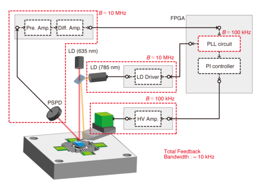
Typical AFM systems consist of hardware including the cantilever probe and scanner unit, and analog circuits to control the system such as the scanner driver. Liquid applications of the AFM require significant improvements in the speed, bandwidth, and sensitivity of detecting the force of probes [1]. “The important feature of the FM-AFM at the WPI-NanoLSI Kanazawa, is that we developed unique digital circuits in order to image solid-liquid interfaces,” explains Miyata. “I developed field programmable gate array (FPGA) digital circuits that enabled information processing in real time. The controller has a bandwidth of 300 kHz to enable ‘frequency shift detection’ [2]. These are examples of the major innovations of the AFM technology that we have developed at Kanazawa.”
Advanced in-liquid FM-AFM research
The FM-AFM systems developed by Miyata and his colleagues at WPI-NanoLSI, Kanazawa are being used to study metal corrosion, lipid layers and microtubules, and hydration structures and dissolution. Recent findings include high speed FM-AFM imaging of calcite (CaCO3, an ionic crystal that is slightly soluble in water) dissolution with image acquisition rates of 2.0 sec/frame (compared with 50 sec/frame for conventional FM-AFM) showing the existence of a transition region indictive of an intermediate state in the dissolution process [3]. The experimental results obtained by FM-AFM measurements were mathematical, atomistic models that simulated the solid-liquid interface.

(Upper figure) High-speed FM-AFM images of calcite dissolution in water. (Lower figure) Molecular dynamics (MD) simulation model of the calcite (10-14) surface in water with a Ca(OH)2 layer at the step edge.
Plans for the FM-AFM at WPI-NanoLSI
“I usually divide my skills into two areas,” says Miyata. “Engineering and AFM. The former means by expertise in the design of field programmable gate array (FPGA), programming with LabVIEW, Python, C, and so on; design of mechanical parts and electrical circuits; and finite element simulations. The latter consists of many years of experience in FM-AFM control for atomic- and molecular-resolution imaging in liquid; in depth knowledge of all the components of an FM-AFM; and analytic skills to compare results from experiments and mathematical simulations. I plan to use all these skills to develop a 3D-AFM platform in order to ‘elucidate the mechanisms of biological phenomena at the atomic and molecular level’, namely the mission of our institute.”
Specific issues to address in the development of high-speed 3D-AFM for biological samples are the necessity for integration with high speed FM-AFM, a new scanner unit and large data recording system, and height position control system.
References
- K. Miyata et al., “Improvements in fundamental performance of liquid-environment atomic force microscopy with true atomic resolution”, Jpn. J. Appl. Phys. 54, 08LA03, (2015).
- K. Miyata et al., “Quantitative comparison of wideband low-latency phase-locked loop circuit designs for high-speed frequency modulation atomic force microscopy”, Beilstein J. Nanotechnol. 9, 1844, (2018).
- K. Miyata et al., “Dissolution Processes at Step Edges of Calcite in Water Investigated by High-speed Frequency Modulation Atomic Force Microscopy and Simulation”, Nano Lett. 17, 4083, (2017).
Further information
https://researchmap.jp/miyata_k (researchmap)


