Unlocking Bio-Molecular Science with HS-AFM
Early Inspirations and Academic Path
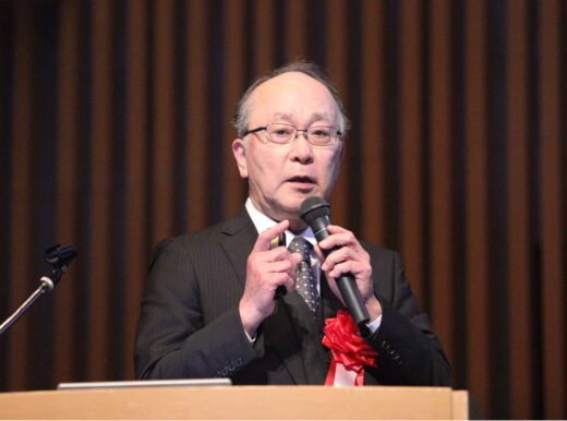
Professor Toshio Ando (Distinguished Professor of Kanazawa University) giving the Commemorative Lecture at the award ceremony for the Leo Esaki Prize on Feb. 26, 2025
Toshio Ando is Professor of Physics and Biophysics at the Nano Life Science Institute (WPI-NanoLSI) at Kanazawa University. Ando’s scientific path was defined early by a love for physics and mathematics, subjects he found more intuitive than memorization-orientated fields like biology and chemistry. As an undergraduate, he initially aspired to study theoretical physics but took experimental biophysics for his graduation project because his preferred professor was on sabbatical. This twist of fate proved decisive. While working on his supervisor’s project, how viscosity affects enzyme activity, he discovered a fascination with experiments. This sparked a lifelong passion for biophysics.
While reflecting on his scientific inspirations, Ando shares that while he respected prominent Japanese physicists like Hideki Yukawa and Shinichiro Tomonaga in his youth, they did not directly influence his career path. His journey was shaped more by personal curiosity than by famous role models.
The Birth of HS-AFM
HS-AFM emerged from Ando’s relentless pursuit of a faster, less intrusive molecular imaging technique. Traditional atomic force microscopy (AFM) was too slow and risked damaging fragile biological samples. To overcome these challenges, Ando designed a novel feedback control system with dynamically adjustable gain—a solution inspired by necessity, not formal training in electronics. This breakthrough allowed HS-AFM to achieve both high speed and precision, revolutionizing molecular biology research.
The journey towards developing HS-AFM was far from straightforward. Ando recalls struggling to balance imaging speed with the delicate nature of biological samples, a challenge that took three years to overcome. He developed a dynamic feedback control circuit that adjusted gain in real-time, reducing errors without damaging samples—a feat that transformed AFM from a static imaging tool into a real-time molecular imaging platform.
Breakthrough Discoveries in Bio-Molecular Dynamics
Ando’s HS-AFM has unlocked insights into fundamental biomolecular functions. His 2010 research challenged conventional thinking about how ATP drives cellular processes, suggesting alternative energy-conversion mechanisms. His team’s real-time imaging of myosin protein molecules moving along actin filaments further demonstrated HS-AFM’s ability to reveal molecular dynamics that other technologies, such as cryo-electron microscopy, struggle to capture.
Ando is still deeply intrigued by ATP’s role as a biological energy source. While conventional wisdom suggests that ATP’s free energy is directly converted into mechanical work, his research points to a more complex mechanism. His studies show that ATP’s energy might drive molecular processes in unexpected ways, reshaping how scientists understand cellular energetics. His team continues to explore how proteins convert ATP’s energy, aiming to reveal principles that could one day inspire artificial molecular machines.
Future Applications of HS-AFM
Looking ahead, Ando predicts HS-AFM will play a crucial role in more complex biological investigations. Current applications focus on purified protein systems, but he envisions extending its use to “in-situ” imaging of biomolecules in their native environments, such as on cell nuclei. He also sees HS-AFM merging with complementary technologies like cryo-electron microscopy to form an integrated system capable of tracking biomolecular dynamics with unparalleled precision. This hybrid approach could redefine how researchers study molecular machinery in action.
He also emphasizes the future potential of combining HS-AFM with time-resolved X-ray crystallography and advanced cryo-electron microscopy. While time-resolved structural methods are emerging, Ando noted that such techniques still need ensemble averaging of molecular structures, making HS-AFM essential for capturing time-specific dynamic molecular events.
Global Scientific Career and Recognition
Ando’s career has been shaped by international experience. Facing limited research funding in Japan, after completing his doctorate he moved to the U.S., where better resources enabled him to collaborate with world-class scientists. This cross-cultural exposure sharpened both his scientific skills and global perspective. Recalling his time in the U.S., he notes the stark contrast in research environments. “U.S. laboratories were well-funded, allowing for dedicated technicians, postdoctoral researchers, and administrative staff—resources that were scarce in Japan,” he says. Returning to Japan, he has thrived, driven by scientific curiosity and multidisciplinary collaboration.
In recognition of his scientific contributions, Ando received the prestigious Medal with Purple Ribbon in 2023 and the Leo Esaki Prize in 2024. Yet, he remains modest, viewing awards as markers of a long research journey rather than endpoints.
Mentorship and Continuing Innovation
As he approaches retirement, Ando is far from slowing down. He plans to publish a second edition of his book on HS-AFM and explore personal projects in programming and electronics—fields he long admired but had little time to pursue.
His advice to aspiring scientists is characteristically bold: “Rebel wisely.” He believes scientific progress often comes from questioning established norms and encourages young researchers to think independently while navigating authority.
Through scientific brilliance and an unwavering drive for discovery, Toshio Ando has left his indelible mark on molecular imaging—unlocking the dynamic bio-molecular world for future generations.
Further information
NanoLSI feature article
High speed in-liquid atomic force microscopy (HS-AFM): Direct visualization of proteins in action
https://nanolsi.kanazawa-u.ac.jp/en/researcher/toshio-ando/
NanoLSI Podcast
Publication of an insightful reference book on high-speed atomic force microscopy (HS-AFM) for in situ biological applications.
https://nanolsi.kanazawa-u.ac.jp/en/researcher/toshio-ando/toshio-ando-page2/
Kanazawa Biophysics Research Team website
https://biophys.w3.kanazawa-u.ac.jp/
Related technical publications
- Ando T., Kodera N., Maruyama D., Takai E., Saito K., and Toda A. “A high-speed atomic force microscope for studying biological macromolecules, Proc. Natl. Acad. Sci. USA98, 12468–12472 (2001).
DOI: 10.1073/pnas.211400898 - Ando T. et al. “High-speed atomic force microscopy for nano-visualization of dynamic biomolecular processes,” Prog. Surf. Sci. 83, 337–437, (2008).
DOI: 10.1016/j.progsurf.2008.09.001 - Kodera N., Yamamoto D., Ishikawa R. and Ando T. “Video imaging of walking myosin V by high-speed atomic force microscopy”, Nature 468, 72–76 (2010).
DOI: 10.1038/nature09450 - Shibata M., Yamashita H., Uchihashi T., Kandori H., and Ando T. “High-speed atomic force microscopy shows dynamic molecular processes in photo-activated bacteriorhodopsin”, Nat. Nanotechnol. 5, 208–212 (2010).
DOI: 10.1038/nnano.2010.7 - Igarashi K., Uchihashi T., Koivula A., Wada M., Kimura S., Okamoto T., Penttilä M., Ando T., and Samejima M. “Traffic jams reduce hydrolytic efficiency of cellulase on cellulose surface”, Science 333, 1279–1282 (2011).
DOI 10.1126/science.1208386 - Uchihashi T., Iino R., Ando T., and Noji H. “High-speed atomic force microscopy reveals rotary catalysis of rotorless F1-ATPase”, Science 333, 755–758 (2011).
DOI: 10.1126/science.1205510 - Uchihashi T., Watanabe Y., Yamasaki T., Watanabe H., Maruno T., Ishii K., Uchiyama S., Song C., Murata K., Iino R., and Ando T. “Dynamic structural states of ClpB involved in its disaggregation function”, Nat. Commun. 9, 2147 (12 pp) (2018).
DOI: 10.1038/s41467-018-04587-w - Kodera, N. et al. “Structural and dynamics analysis of intrinsically disordered proteins by high-speed atomic force microscopy”, Nat. Nanotechnol. 16, 181-189 (2021).
DOI: 10.1038/s41565-020-00798-9
Book
Ando, Toshio. High-Speed Atomic Force Microscopy in Biology: Directly Watching Dynamics of Biomolecules in Action. 1st ed., Springer, March 2022. ISBN: 978-3-662-64783-7 (Print), 978-3-662-64785-1 (Electronic).
DOI: 10.1007/978-3-662-64785-1
https://link.springer.com/book/10.1007/978-3-662-64785-1
Posted: January, 2025