High-speed atomic force microscopy: In-situ and real-time visualization of biomolecular dynamics of myosin
High-speed atomic force microscope (HS-AFM) imaging of myosin V yielded unprecedented insights into the dynamics of molecular cargo transporter in cells.
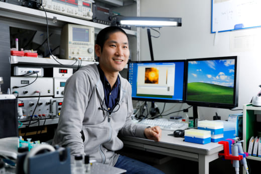
古寺 哲幸、金沢大学ナノ生命科学研究所 教授
The HS-AFM technology at Kanazawa University exhibits the highest performance in the world with a frame rate of 30 ms/frame, a tapping force of ~30 pN, and spatial resolution of ~1 nm.
Noriyuki Kodera has been one of the key players in the development of the HS-AFM and was awarded the prestigious 14th Japan Society for the Promotion of Science (JSPS) Prize for his contributions to AFM imaging [1]. “My expertise is in improvement of scanning performance and functional extension of the HS-AFM system developed by Professor Toshio Ando of Kanazawa University, and in visualizing biological samples with the HS-AFM system”, says Kodera. “I have been working with Professor Ando since my Bachelor’s degree. The research on visualizing myosin at work was extremely challenging, taking many years of perseverance before we succeeded in obtaining a movie in 2010. Our results were published in Nature”. The movie clearly showed that a double-headed myosin-V molecule is walking along an actin filament [1].
The HS-AFM, born at Kanazawa University, is a tapping mode system that is now commercially available and used worldwide for imaging biological samples at work. Importantly, a special feedback control maintains an interaction force between the cantilever tip and sample surface constant with a wide bandwidth. Furthermore, the short 7 micrometer long cantilever with a small spring constant (compared with approximately 200 micrometers in conventional AFMs) enables much faster and very gentle scans (Fig. 1).
“The HS-AFM enabled us to perform real time imaging of functioning biomolecules at nano-meter and sub-second spatiotemporal resolution, as shown in movies posted on our website,” says Kodera. “The performance of our HS-AFM is the best in the world for studying the dynamic behaviors of biomolecules. In addition to myosin V, we have also directly visualized antigen-antibody interactions, unidirectional binding of cofilin (an actin depolymerizing protein) to actin filament, and more recently the genome editing action by CRISPR-Cas9 reported by Mikihiro Shibata.” [2]
Kodera and his colleagues have also visualized the thin and flexible structure of “intrinsically disordered proteins”. “This means HS-AFM can directly visualize a wobbly moving single polypeptide chain,” explains Kodera. “These direct observations yield both visual insights into the dynamic behaviors of functioning proteins and importantly, shed light on their functional mechanisms.”
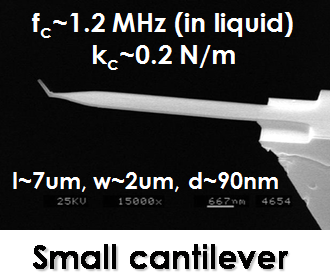
Fig. 1: Small cantilever for HS-AFM system
Research highlights
Noriyuki Kodera put HS-AFM on the world stage with the publication of the first direct visualization of myosin V molecules translocating along actin filaments. This report gave direct visual confirmation of molecular dynamics such as lever-arm swing, foot stomping, unfolding of the coiled-coil tail, and so on. This paper opened up many new interdisciplinary areas of research based on HS-AFM imaging of ‘functional biomolecules’.

Myosin V walking along actin filament
参考文献
- N. Kodera, D. Yamamoto, R. Ishikawa & T. Ando, “Video imaging of walking myosin V by high-speed atomic force microscopy”, Nature 468, 72–76, (2010). doi:10.1038/nature09450
- M. Shibata, H. Nishimasu, N. Kodera, S. Hirano, T. Ando, T. Uchihashi & O. Nureki, “Real-space and real-time dynamics of CRISPR-Cas9 visualized by high-speed atomic force microscopy”, Nature Communications 8, 1430, (2017). doi:10.1038/s41467-017-01466-8