High speed non-invasive cell imaging and electrochemical activity mapping
Marina Makarova, Assistant Professor
“My background is materials science,” says Marina Makarova, an assistant professor at the Nano Life Science Institute, Kanazawa University (WPI-NanoLSI). “After completing my doctorate at the Faculty of Materials Sciences of Moscow State University in 2003, I moved to the Solid State Electrocatalysis Group at the J. Heyrovsky Institute of Physical Chemistry, Prague, Czech Republic. Then, I moved to the Group of Optical and Biophysical systems at the Institute of Physics CAS also in Prague. Between 2011 and 2014 I joined the National Institute of Materials Sciences (NIMS), Tsukuba, as a MANA Research Associate under the supervision of Dr. Yuji Okawa (deceased in 2019), who became my teacher of Scanning Probe Microscopy. After this I returned to the Institute of Physics CAS, Prague, Czech Republic and then joined Akita University for approximately 2 years. And I joined the NanoLSI in December 2018. Quite a busy time!”
Makarova’s dedication to her research is reflected by the awards that she has received including several ‘George Soros Foundation International Scholarships of Soros Science Education Program’ between 1995-2000 and Makarova was a finalist in the ‘L’Oreal Program Grant for Young Female Scientists’ in 2010.
Making plans for spearhead nanopore sensors
After joining the NanoLSI Makarova has started combining her expertise in materials science with the world-class scanning probe microscopy instruments at the NanoSLI for high speed and non-invasive imaging of living cells to gain insights into the onset and diagnosis of cancer.
“My plans include developing stable ‘spearhead nanopore sensors’ for mapping fluctuations in pH, cation or anion in multifunctional cell imaging,” explains Makarova. “This is because elements such as calcium, iron, and reactive nitrogen or oxygen species participate in biochemical processes including cancer formation.”
Makarova is also developing sensors based on the electrical conductivity measurements of long-chain molecules. Examples include field-effect and organic electrochemical transistors. The aim is to create a double probe platform to directly measure electrical current passing through single molecules of analyte and potentially detect early stages of cancer disease or even sequence DNA. Another idea is to use 1D polymer nanowires between sealed carbon electrodes.
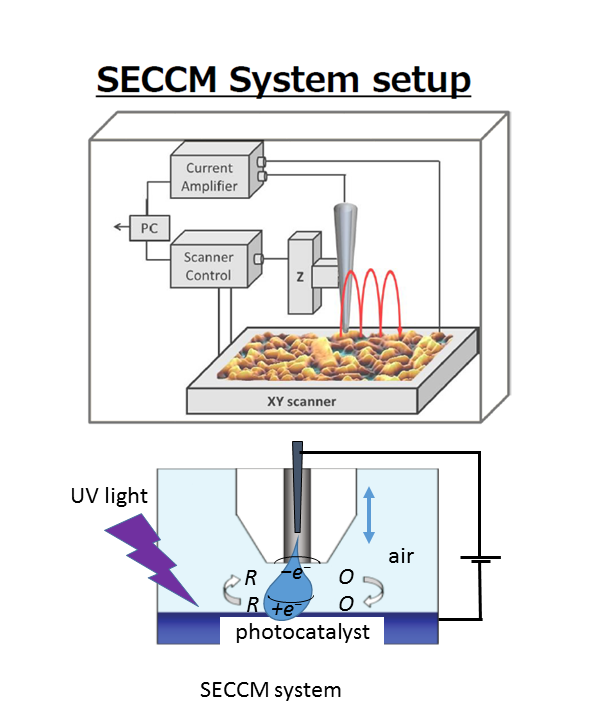
The initial focus is on the development of Scanning Ion Conductance Microscopy (SICM) for cell imaging and its derivative Scanning Electrochemical Cell Microscopy (SECCM) for electrochemical activity mapping. Specifically, ion currents are monitored for topographic information without damaging the surfaces of samples. SECCM provides the way to map electrocatalytic activity of nanostructures to study the processes of energy conversion such as water splitting and photosynthesis. Both methods use laser-pulled nanopipettes with 50-100 nm hole diameters and pipettes could be functionalized for sensor measurements, with double barrel pipettes encapsulate with carbon electrodes to measure current at about 10 nm above specimens.
“Currently I am making concrete plans for my research,” says Makarova. “For example, I am thinking about functionalizing the insides of nano pipettes with inorganic oxide layers to increase stability and improved chemisorption with high affinity metal ion such as Ca2+ or Na+ that would act as a biomarker.”
Makarova stresses that it will be difficult to form oxide layers inside nano pipettes using conventional methods that necessitate high temperatures, alkaline conditions, and the possibility of clogging of pipette orifices. “I am going to experiment with sol-gel formation of crystalline nanoparticles at room temperature to overcome the limitations of conventional approaches.”
Mastering the Japanese language
Plans include (1) developing a double-probe platform with the nanometer gap between electrodes to characterize single-chain (bio)molecules and sequence DNA; and (2) studying of photoeffects such as photosynthesisor plasmonic resonance on electrocatalysis and hydrogen evolution by SECCM microscopy.
“The NanoLSI is a great environment for conducting research,” says Makarova. “The administration staff and other scientists provide great support for my research. People sometimes ask if there what is it like to work as a female researcher. The answer is that I have not had to think about such things. I have nothing but admiration for the way the NanoLSI is structured for everyone’s needs, whether they are female, male, from overseas or Japan. The support is excellent. One of my personal goals is to master the Japanese language. So, I am taking a lecture course given by the university. I am looking forward to contributing to the research at the NanoLSI.”
References
- M. V. Makarova, Y. Akaishi, T. Ikarashi, K. S. Rao, S. Yoshimura, H. Saito, “Alternating Magnetic Force Microscopy: Effect of Si doping on the temporal performance degradation of amorphous FeCoB magnetic tips”, Journal of Magnetism and Magnetic Materials 471 209 – 214, (2019).
https://doi.org/10.1016/j.jmmm.2018.09.046
- Marina Makarova et al “Self-assembled diacetylene molecular wire polymerization on an insulating hexagonal boron nitride (0001) surface”, Nanotechnology 27 395303, (2016).
https://doi.org/10.1088/0957-4484/27/39/395303
- Marina Makarova, Swapan K. Mandal, Yuji Okawa, Masakazu Aono, “Ordered Monomolecular Layers as a Template for the Regular Arrangement of Gold Nanoparticles”, Langmuir 29, 24, 7334-7343, (2013).
https://doi.org/10.1021/la400177u
Further information
Marina Makarova
https://www.fzu.cz/en
Posted May 18,2020