[For Internal Applicants Only] NanoLSI–Nikon Solutions Joint Workshop has been postponed
Important Notice
Aug. 19, 2021, regrettably we decided to postpone NanoLSI–Nikon Solutions Joint Workshop due to the COVID-19 infection spread. The details will be sent to registrants by e-mail. Please check.
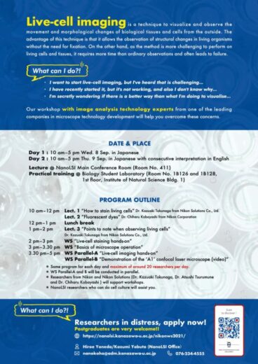
Researchers in distress, apply now!! Postgraduates are very welcome!!

- I want to start live-cell imaging, but I’ve heard that is challenging…
- I have recently started it, but it’s not working, and also I don’t know why…
- I’m secretly wondering if there is a better way than what I’m doing to visualize…
Our workshop..
with image analysis technology experts from one of the leading companies in microscope technology development will help you overcome these concerns.
Live-cell imaging..
is a technique to visualize and observe the movement and morphological changes of biological tissues and cells from the outside. The advantage of this technique is that it allows the observation of structural changes in living organisms without the need for fixation. On the other hand, as the method is more challenging to perform on living cells and tissues, it requires more time than ordinary observation and often leads to failure.
DATE & PLACE
Day 1 : 10 am–5 pm Wed. 8 Sep. in Japanese
Day 2 : 10 am–5 pm Thu. 9 Sep. in Japanese with consecutive interpretation in English
Lecture @ NanoLSI Main Conference Room (Room No. 411)
Practical training @ Biology Student Laboratory (Room No. 1B126 and 1B128, 1st floor, Institute of Natural Science Bldg. 1)
PROGRAM OUTLINE
| 10.00 am–12.00 pm | Lect. 1 “How to stain a living cells” Dr. Kazuaki Tokunaga from Nikon Solutions Co., Ltd. Lect. 2 “Fluorescent dyes” Dr. Chiharu Kobayashi from Nikon Corporation |
| 12.00 pm-1.00 pm | Lunch break |
| 1.00 pm – 2.00 pm | Lect. 3 “Points to notes when observing living cell” Dr.Kazuaki Tokunaga from Nikon Solutions Co., Ltd. |
| 2.00 pm – 3.00 pm | WS “Live-cell staining hands-on” |
| 3.00 pm – 3.30 pm | WS “Basic of microscope operation” |
| 3.30 pm – 5.00 pm | WS Parallel-A “Living Cell Imaging Hands-on” WS Parallel-B “Demonstration of the ‘A1’ confocal laser microscope (video)” |
- Same program for each day and maximum of around 20 participants per day.
- WS Parallel-A and B will be conducted in parallel.
- Researchers from Nikon and Nikon Solutions ( Dr. Kazuaki Tokunaga, Dr. Atsushi Tsurumune and Dr. Chiharu Kobayashi ) will support workshops.
- NanoLSI researchers who can do cell culture will assist you.
You’re ready to apply!
Google Form ”Registration required in advance.”
https://forms.gle/HBN75S42x9zx72Js6
***Please note that we will cease accepting applications once the limit is reached.
***We may decide to postpone or cancel this workshop depending on the situation of the spread of the COVID-19. We appreciate your understanding.

👤 Hiroe Yoneda, Kazumi Yabuta (NanoLSI Office)
📞 (076) 234-4555
📧nanokoho[at]adm.kanazawa-u.ac.jp Please replace [at] with @.

